Infrastructure
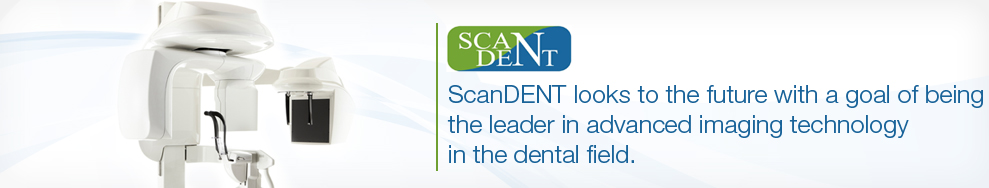
TechnologyCone Beam Computed Tomography (CBCT) is a proven recent advancement in the field of Dento Maxillo Facial Imaging. As compared to Multidetector Computed Tomography (MDCT) CBCT generates 3D images at a higher speed, lower absorbed dose of radiation and at economical cost.
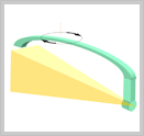
CBCT makes use of a Cone Shaped X ray beam and a TFT detector which move in tandem around the patient's head to take a single scan covering the entire region of interest in a matter of a few seconds. This leads to a considerable reduction in the amount of absorbed dose of radiation to the patient, operating personnel and the environment.
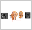
CBCT produces panoramic and cephalometric projections, which become 3-D when the data is reformatted in a volume. The images that result can be manipulated with the machine's visualization software from any point of view: in the axial, coronal, saggital and cross-sectional planes.
Layers can be "peeled away" to observe underlying anatomical structures and defects, slice thickness can be modified to suit individual requirements, areas of interest can be zoomed and highlighted, measurements can be performed between various structures. Diagnosis, treatment decisions and thereby the prognosis can benefit from these advanced views of the maxillofacial complex.
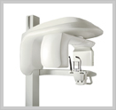
Image contrast and ability to visualize soft tissues still remains a challenge. However our CBCT units have multiple FOV's, higher resolution, lower radiation exposure and are less expensive. They are exclusively designed for dental applications.